
- 面议
起订量:
Cell volume measurement Cell volume assay实时跟踪贴壁细胞体积
- 型号
- Cell volume measurement

该企业相似产品


世联博研(北京)科技有限公司(Bio Excellence International Tech Co.,Ltd)简称为世联博研。世联博研是一家集进口科研仪器代理销售以及实验技术服务于一体的技术公司。世联博研专注生物力学和3D生物打印前沿科研设备代理销售及科研实验项目合作服务,内容涵盖了血管力学生物学、生物力学建模仿真与应用、细胞分子生物力学、组织修复生物力学、骨与关节生物力学、口腔力学生物学、眼耳鼻咽喉生物力学、康复工程生物力学、生物材料力学与仿生学、人体运动生物力学等生物力学研究以及生物材料打印、打印样品生物力学性能测试分析的前沿领域科研利器和科研服务。
世联博研的客户范围:
科研院所单位、生物医学科研高校、医院基础科研单位等。
世联博研公司代理的品牌具有:
1)近10年长期稳定的货源
2)以生物力学、细胞力学、细胞生物分子学、生物医学组织工程、生物材料学为主,兼顾其他相关产品线
3)提供专业产品培训和销售培训
4)良好的技术支持
5)已成交老客户考证
6)每年新增的货源。
详细信息
Cell volume assay实时跟踪贴壁细胞体积
Cell volume assay
Tracking the cell volume of adherent cells in real time
4DCELL DEVICE
Cell volume measurement technology
READ-OUTS
Cell volume, ion pumps
STANDARD CULTURE LIMITATION
There are several physiological and pathological processes where cells undergo a change of volume. However, there are no reliable methods that can be applied to accurately measure volume of adherent cells in real time.
CELL VOLUME ASSAY BENEFITS
Cells are cultured in an optically transparent chamber that enables to accurately determine their volume along time and to follow in parallel the biochemical processes responsible for the volume change, as for example activation of ion pumps.
EXAMPLES
Volume tracking from interphase stage, to mitosis of Raji cells [2].
![]()
(A) Cells are placed in poly(dimethylsiloxan) chambers of calibrated height set by pillars, in medium supplemented with FITC-Dextran. Bottom picture: cells exclude fluorescence on epifluorescence images (Scale bar 20 mm).
(B) The fluorescence profile corresponding to the dotted line in (A): maximum and minimum of fluorescence intensity correspond to chamber maximal height (background) and zero height (pillar), respectively. Right: these values are used to calibrate the signal and calculate the optical thickness of the cells.
(C) Finally, cell volume is obtained by integrating the total fluorescence intensity over the cell area.
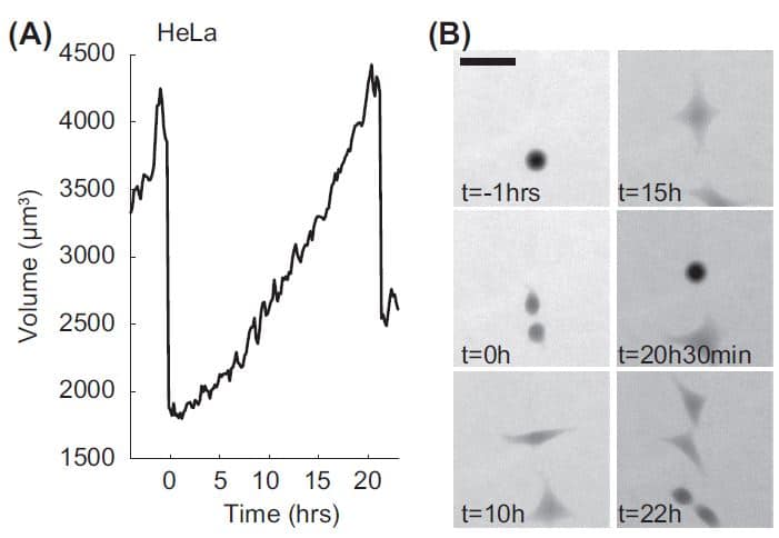
(A) Volume trajectory of a HeLa cell. The two volume overshoots at the beginning and the end correspond to transient volume increase in mitosis with the first one corresponding to the mother cell and the second one to the daughter cell.
(B) Raw Fluorescence images of the cell in (A) with FXm. Scale bar 50 mm.
REFERENCES
[1] Zlotek-Zlotkiewicz, E. et al. (2015). Journal of Cell Biology, 211(4), 765–774.
[2] Cadart, C et al. (2017). Methods in Cell Biology, 139,103-120.


