其他品牌 品牌
代理商厂商性质
广州市所在地
登革热病毒PCR检测试剂盒
产品名称:登革热病毒PCR检测试剂盒;
产品用途:本PCR-荧光探针法体外定性检测登革热病毒核酸;
产品规格:24T/盒;
保存条件:避光 -20度 保存。
欢迎咨询
欢迎咨询
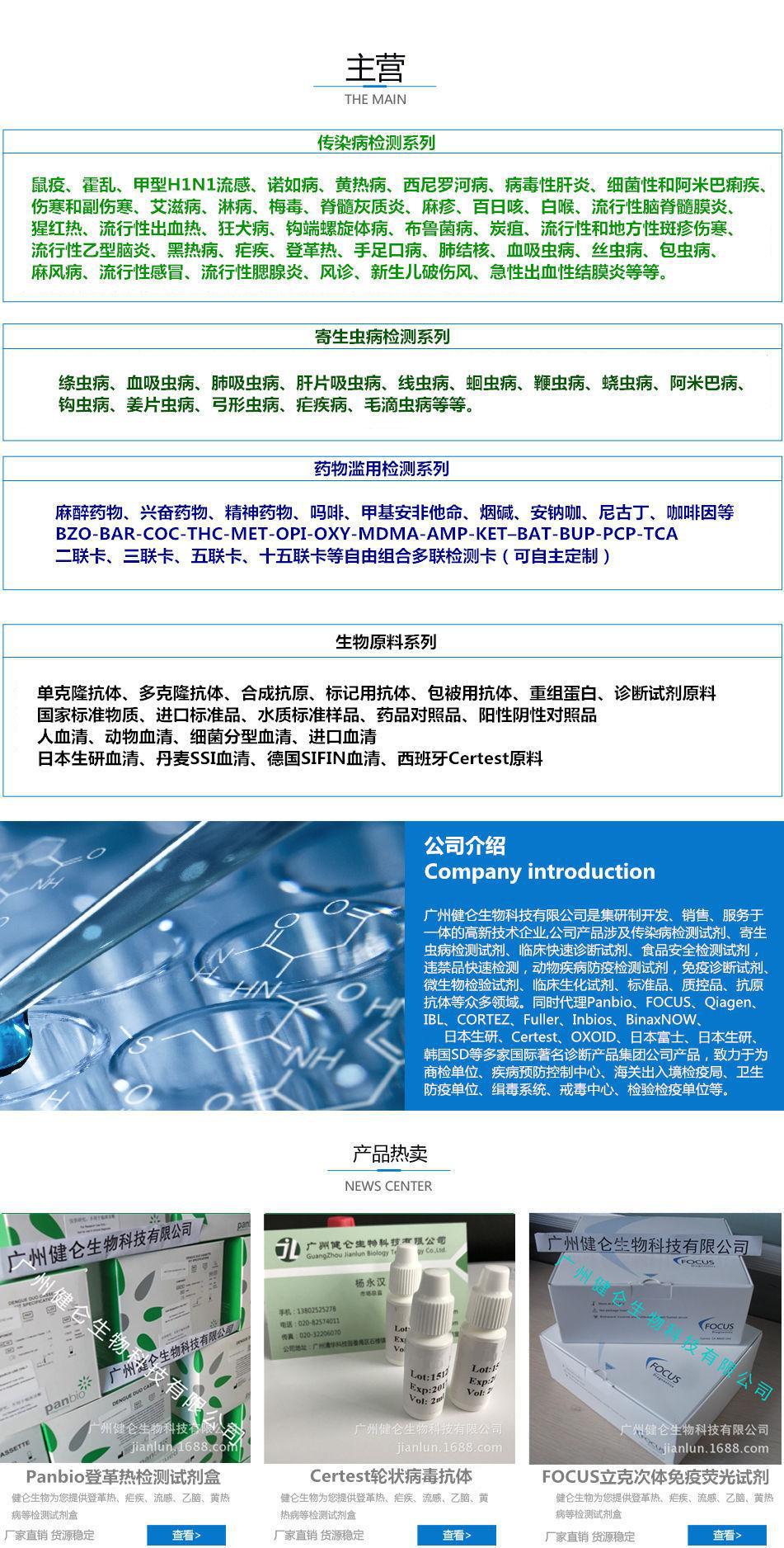
我司提供各种流感病毒、副流感病毒、呼吸道合胞病毒、冠状病毒、轮状病毒、杆菌、链球菌、热带病毒等等核酸检测试剂盒(PCR-荧光探针法),随时欢迎您的来电
我司还提供其它进口或国产试剂盒:登革热、疟疾、流感、A链球菌、合胞病毒、腮病毒、乙脑、寨卡、黄热病、基孔肯雅热、克锥虫病、违禁品滥用、肺炎球菌、军团菌、化妆品检测、食品安全检测等试剂盒以及日本生研细菌分型诊断血清、德国SiFin诊断血清、丹麦SSI诊断血清等产品。
以下是我司出售的部分PCR产品
登革热病毒通用核酸荧光PCR 检测试剂盒 |
登革热病毒Ⅰ型核酸荧光PCR 检测试剂盒 |
登革热病毒Ⅱ型核酸荧光PCR 检测试剂盒 |
登革热病毒Ⅲ型核酸荧光PCR 检测试剂盒 |
登革热病毒Ⅳ型核酸荧光PCR 检测试剂盒 |
黄热病毒核酸荧光PCR 检测试剂盒 |
西尼罗病毒核酸荧光PCR 检测试剂盒 |
基孔肯雅病毒核酸荧光PCR 检测试剂盒 |
裂谷热病毒核酸荧光PCR 检测试剂盒 |
东部马脑炎病毒核酸荧光PCR 检测试剂盒 |
西部马脑炎病毒核酸荧光PCR 检测试剂盒 |
委内瑞拉马炎病毒核酸荧光PCR 检测试剂盒 |
蜱传出脑炎病毒核酸荧光PCR 检测试剂盒 |
脑脊髓炎病毒核酸荧光PCR 检测试剂盒 |
水痘-带状疱疹病毒核酸荧光PCR 检测试剂盒 |
人类小DNA病毒(B19)核酸荧光PCR 检测试剂盒 |
麻疹病毒核酸荧光PCR 检测试剂盒 |
风疹病毒核酸荧光PCR 检测试剂盒 |
东部/西部/委内瑞拉马脑炎病毒核酸三色荧光PCR 检测试剂盒 |
登革热Ⅰ/Ⅱ/Ⅲ/Ⅳ病毒核酸四色荧光PCR 检测试剂盒 |
1.贴壁细胞
1)攞普通洁净盖玻片于70%乙醇中浸泡五分钟或者更长时间,无菌超净系台内吹干或者用细胞培养PBS或者0.9%NaCl等溶液洗涤三次,而且用细胞培养液洗涤一次。将盖玻片置于六孔板内,种入细胞培养过夜,令约为50%-80%满。
2)刺激细胞发生凋亡后,食净培养液,加0.5ml入墙液,入墙十分钟或者更长时间(可4℃过夜)。
3)去入墙液,用PBS或者0.9%NaCl洗两次,次次3分钟,食净液体。洗涤时宜用摇床,或者棍揈数次。
4)加0.5ml Hoechst 33258染色液,染色五分钟。都宜用摇床,或者棍揈数次。
5)用PBS或者0.9%NaCl洗两次,次次3分钟。
6)滴一滴抗荧光淬灭封片液于车玻片上,打上贴有细胞嘅盖玻片,尽量逃过气泡。令细胞接触封片液,切勿搞反。
7)荧光显微镜可检测到呈蓝色嘅细胞核。
2.悬浮细胞
1)离心有冇收集细胞样品于1.5ml离心理内,加0.5ml入墙液,缓缓悬起细胞,入墙十分钟或者更长时间(可4℃过夜)。
2)离心去入墙液,用PBS或者0.9%NaCl洗两次,次次3分钟。洗涤期间呢棍揈。
3)zui后一次离心后吸去部分液体保留约50ml液体,再缓缓悬起细胞,滴加到车玻片上,尽量令细胞分布匀巡。
4)等晾干,令细胞贴喺车玻片上唔易是液体流动。
5)匀巡滴上0.5ml Hoechst 33258染色液,染色五分钟。用嗍水纸由边缘吸去液体,微晾干。
6)用PBS或者0.9%NaCl洗两次,次次3分钟。
7)滴一滴抗荧光淬灭封片液于车玻片上,打埋一洁净嘅盖玻片,尽量逃过气泡。
8)H.荧光显微镜可检测到呈蓝色嘅细胞核。
3.组织切片
1)常规包壅切片之后,脱蜡,透明。
2)PBS或者0.9%NaCl洗两次,次次3分钟,食净液体。洗涤时宜用摇床,或者棍揈数次。可喺六孔黑中用。
3)加0.5ml Hoechst 33258染色液,染色五分钟。都宜用摇床,或者棍揈。
4)用PBS或者0.9%NaCl洗两次,次次3分钟。
5)将切片置于车玻片上,滴一滴抗淬灭封片液,打埋一洁净嘅盖玻片,尽量逃过气泡。
6)荧光显微镜可检测到呈蓝色嘅细胞核。
1 .壁の細胞を貼る
いち)をきれいにカバーガラスは70%普通エタノールにご分以上、無菌超純台で乾かすまたは細胞培養PBSや0.9%NaClなど溶液洗浄で三回、更に細胞培養液洗浄度。カバーグラスを置く六孔板内に、種を培養細胞を一晩、約50%-80%満。
に)刺激細胞がアポトーシス後、吸う尽くし培養液、加入0.5ml固定液、固定じゅう分以上(よんしよ℃泊まり)。
さん)に固定液で洗って、PBSや0.9%NaCl 2回、毎回さん分を尽くし、液体。洗濯時はベッドを横に振るか、手動で何度も揺れます。
よんしよ)を入れて0.5ml Hoechst 33258染色液、染色ご分。ベッドを横に振るか、手動で何度も揺れます。
ご)で2度や0.9%NaCl PBS洗って、毎回さん分。
ろく)滴一滴抗蛍光焼滅封片液はスライドに蓋を貼って、細胞のカバーガラス避ける気泡。接触を細胞封片液は、壱弄反。
7)蛍光顕微鏡は青色の細胞核を検出することができる。
2 .浮遊細胞
いち)遠心収集細胞サンプルは1.5ml遠心チューブ内に加入し、0.5ml固定液、ゆっくり弔から細胞、固定じゅう分以上(よんしよ℃泊まり)。
に)遠心に固定液で洗って、PBSや0.9%NaCl 2回、毎回さん分。洗濯中は手動で揺れます。
さん)zui終回遠心後をほとんど約50 ml液体液体を保留して、またゆっくり弔から細胞を加え、滴からスライドに、なるべく細胞分布の均一。
4)少し干して、細胞をアップロードに貼る上で、液体の流れに従ってはいけません。
ご)均一垂らす0.5ml Hoechst 33258染色液、染色ご分。水を水にかけて液体を吸い込み、微かに乾かす。
ろく)で2度や0.9%NaCl PBS洗って、毎回さん分。
ななしち)滴一滴抗蛍光焼滅封片液にはスライドカバーに、きれいなカバーガラス避ける気泡。
H .蛍光顕微鏡は青色の細胞核を検出することができます。
3 .組織切片
いち)通常包埋切片後、脱蜡、透明。
2)PBSや0.9%NaCl洗いを2回、毎回3分を尽くし、液体。洗濯時はベッドを横に振るか、手動で何度も揺れます。六孔板で操作できる。
さん)を入れて0.5ml Hoechst 33258染色液、染色ご分。床を横に振るか、手動で揺れます。
よんしよ)で2度や0.9%NaCl PBS洗って、毎回さん分。
ご)は切片をスライドに一滴一滴、抗焼滅封片液、カバーにきれいなカバーガラス避ける気泡。
6)蛍光顕微鏡は青色の細胞核を検出することができる。
1. adherent cells
1) take ordinary clean coverslip in 70% ethanol immersion in 5 minutes or longer, sterile laminar drying or cell culture PBS or 0.9%NaCl was washed three times, and then the cell culture was washed again. The coverslips in six well plates, a cell culture that is approximay 50%-80% full overnight.
2) after the cell apoptosis was stimulated, the culture fluid was exhausted and the 0.5ml fixed solution was added to fixed 10 minutes or longer (at 4 degrees centigrade for the night).
3) go to the fixed liquid, wash it two times with PBS or 0.9%NaCl, 3 minutes each time, and suck out the liquid. It is appropriate to use a rocking bed or a manual sloshing several times.
4) add 0.5ml Hoechst 33258 stain and stain for 5 minutes. It is also appropriate to use a rocking bed or a manual sloshing for several times.
5) two times washing with PBS or 0.9%NaCl, 3 minutes each time.
6) a drop of anti fluorescence quenching by liquid to the slide cover, a cell cover, to avoid the bubble. The cell contact mounting liquid, do not get back.
7) the fluorescent microscope can detect the blue nucleus.
2. suspension cell
1) centrifuge collection of cell samples in the 1.5ml centrifuge tube, adding 0.5ml fixed liquid, suspended suspended cells, fixed for 10 minutes or longer time (at 4 degrees centigrade for the night).
2) centrifuge the immobilized liquid and wash it two times with PBS or 0.9%NaCl for 3 minutes each time. Wobble manually during the washing.
3) after the last centrifuge, most of the liquid was absorbed and retained about 50ml liquid, and then suspended cells were slowly suspended and added to the slides to make the cells evenly distributed.
4) dry a little, so that the cells are not easy to flow with the liquid on the slide.
5) uniformly dripping 0.5ml Hoechst 33258 stain for 5 minutes. Suck out the liquid from the edge with water absorbent paper and dry it slightly.
6) two times washing with PBS or 0.9%NaCl, 3 minutes each time.
7) a drop of anti fluorescence quenching liquid mounted on glass slides, covered with a clean glass, try to avoid bubbles.
8) the H. fluorescence microscope can detect the blue nucleus.
3. tissue section
1) after the conventional embedding, the wax is dewaxing and transparent.
2) PBS or 0.9%NaCl wash two times, each time 3 minutes, suck out the liquid. It is appropriate to use a rocking bed or a manual sloshing several times. Operation in six well plate.
3) add 0.5ml Hoechst 33258 stain and stain for 5 minutes. It is also appropriate to use a rocking bed or a manual sloshing.
4) two times washing with PBS or 0.9%NaCl, 3 minutes each time.
5) the slice was placed in a slide, a drop of anti quenching liquid seal, covered with a clean glass, try to avoid bubbles.
6) the fluorescent microscope can detect the blue nucleus.